
From a Protein Sequence to a Predicted Structure: Running a BioLM Job End to End

For many researchers, running an artificial intelligence-driven protein structure prediction job is treated as a computational black box. The practitioner inputs a raw amino acid sequence, waits for a progress bar to compile, and downloads a resulting PDB or CIF coordinate file. What happens during that execution window, or how to systematically evaluate the physical coordinate outputs before trusting them with downstream wet-lab resources, remains poorly understood.
This technical guide demystifies this execution pipeline, walking through the end-to-end process of running a structure prediction job using Purna’s BioLM Jobs workspace. We outline the mechanics of sequence input preprocessing, the process of model selection on the BioCompute runtime, how to read output confidence metrics, and the precise biophysical checks required to validate the resulting coordinates.
The BioLM Job Execution Pipeline
The pipeline moves sequentially from raw, unmapped character strings to a validated, coordinate-level structural representation:
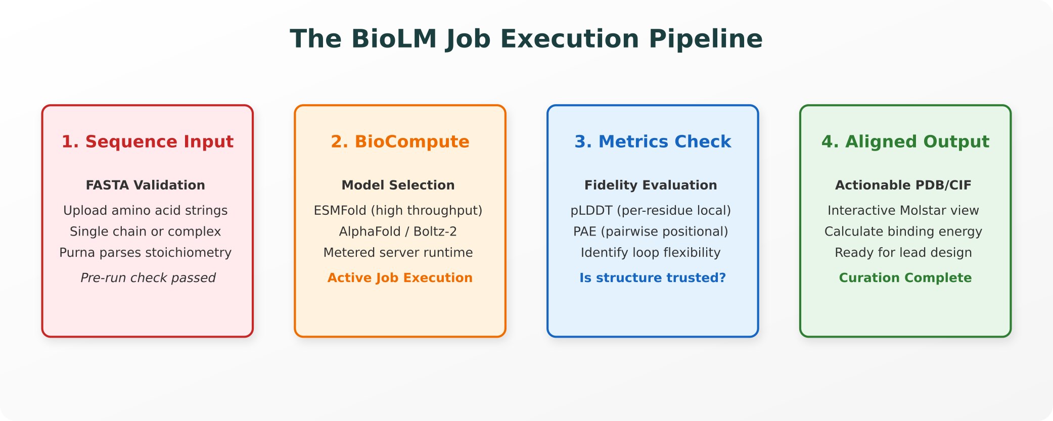
1. The Starting Point: Input Sequence Preprocessing
Every structure prediction job begins with a raw character string representing the protein sequence, standardly structured in FASTA format.
- Single-Chain vs. Complexes: For a single, monomeric protein, you input a single, continuous string of standard amino acid letters. If you are predicting a multimeric complex or protein-protein interface, the sequences must be formatted as distinct chains separated by a colon or submitted with defined stoichiometry in your run parameters.
- Validation and Cleaning: Before a job enters the active queue, Purna’s preprocessing engine runs a validation check. It automatically strips non-standard or degenerate amino acid characters (such as X, B, or Z), flags duplicated sequence headers, and checks the sequence length. Length checks are highly critical: while high-throughput models can handle small domains in seconds, predicting large, multi-chain complexes containing thousands of residues requires substantial GPU memory allocation.
2. Running the Job: Model Selection and Runtime Execution
Once the input sequence passes validation, the researcher must select the appropriate structural model on Purna’s BioCompute runtime:
- High-Throughput Curation (ESMFold): If you are running an active variant scan or screening hundreds of candidate mutant sequences, ESMFold is the standard choice. It bypasses the slow Multiple Sequence Alignment (MSA) step by utilizing a massive transformer-based protein language model, predicting single-chain structures in seconds directly from sequence letters.
- Complex Folding and Multi-Domain Targets (AlphaFold3 / Boltz-2): If your target involves a complex multimeric interface, structural nucleic acids (DNA/RNA), or ligand-binding interactions, you select AlphaFold3 or Boltz-2. These models generate MSA profiles by searching global sequence databases, utilizing diffusion-based architectures to predict complex, multi-component structural coordinates.
- Designing De Novo Scaffolds (RFdiffusion): If the objective is to generate an entirely new, non-natural protein binder to occupy a target active site, the workflow transitions from prediction to design, calling RFdiffusion pipelines to generate structural backbones from scratch.
During this execution window, Purna’s BioCompute runtime allocates specialized, high-performance GPU instances, manages memory requirements, and compiles the raw coordinate outputs.
3. Reading the Output: Understanding Confidence Metrics
When a BioLM Job completes, the output contains more than just a 3D coordinate model. It delivers a comprehensive set of statistical confidence scores that must be evaluated:
pLDDT (predicted Local Distance Difference Test)
pLDDT is a per-residue local confidence score ranging from 0 to 100, plotted as a continuous graph along the protein’s sequence backbone.
- 90 to 100 (Very High): Indicates high structural fidelity. The predicted side-chain coordinates are highly accurate, and the structure can be trusted for rational drug design or active-site modeling.
- 70 to 90 (Good): Indicates a reliable backbone fold, suitable for general structural biology and domain mapping.
- 50 to 70 (Low): Suggests some structural flexibility. These regions should be interpreted with caution.
- Below 50 (Very Low): Strongly correlates with intrinsically disordered or unstructured regions.
PAE (Predicted Aligned Error)
PAE is a 2D distance-error matrix that measures pairwise positional confidence. It plots the predicted distance error (in Angstroms) between every pair of residues in the protein.
- The Interpretation: If residue A is correctly positioned relative to residue B, the PAE at coordinate (A, B) will be low (dark blue).
- Why It Matters: While pLDDT tells you if individual domains are folded correctly, PAE is the only metric that tells you if those domains are correctly positioned relative to each other. A multimeric interface or multi-domain linker with high pLDDT but high PAE indicates that the individual components are folded, but their relative orientation is completely uncertain.
4. What to Check Before Trusting the Result
To maintain rigorous scientific standards, computational biologists should run several quality control checks before advancing a predicted structure to experimental screening:
- Identify Unstructured Loops: Check for segments with pLDDT scores below 50. These typically represent highly flexible, disordered regions. If these loops occupy your target binding interface, the predicted docking pose is highly likely to be biologically irrelevant.
- Evaluate Interface PAE: If you are analyzing a protein-protein complex, inspect the interface region on your PAE plot. A high-error boundary at the interface indicates a low-confidence docking prediction, meaning the complex orientation cannot be trusted.
- Check Training Data Homology: If your sequence has close structural homologs in the Protein Data Bank (PDB), the prediction is highly likely to be extremely accurate. However, if your sequence represents a completely novel fold with no known homologs in the training dataset, the model’s prediction represents a statistical extrapolation that requires extensive wet-lab validation.
Closing: Calibrated Epistemic Trust
The true deliverable of a Purna BioLM Job is not simply a static 3D PDB file. Rather, it is the structural coordinate model combined with a calibrated, scientifically precise understanding of where the model is highly confident and where it remains speculative.
By utilizing pLDDT and PAE plots to map structural confidence, researchers can ensure they apply their expert judgment and wet-lab validation resources to the specific regions that require experimental confirmation, transforming structure prediction from a black-box guessing game into a precise, evidence-grounded scientific tool.
Explore Purna's Molecular Intelligence Platform
AI-powered workspace for biology teams to accelerate drug discovery from target identification to lead optimization.
Try Purna AI →
