
What Codon Optimization Actually Changes in Your Construct, and How to Check It

For many molecular biologists, designing a synthetic gene construct involves a standard, black-box routine. The researcher pastes their wild-type sequence into a vendor’s online portal, clicks “optimize for host,” and adds the resulting sequence to their ordering cart. Because the vendor’s algorithms handle the sequence re-alignment, codon optimization is frequently treated as a simple pre-synthesis checkbox.
However, clicking that button introduces extensive, highly consequential changes to the physical sequence of your construct. While the encoded amino acid sequence remains fixed by design, the nucleotide sequence, secondary structures, and GC content distribution are completely rewritten. Treating this process as a black box without performing independent human verification regularly leads to synthesis failures, poor expression yields, or lost biological functions.
This article serves as a definitive, technically precise reference guide to codon optimization. We examine the specific biophysical mechanics changed during an optimization run, discuss what algorithmic tools do and do not guarantee, and outline a concrete verification checklist researchers should run before placing a synthesis order.
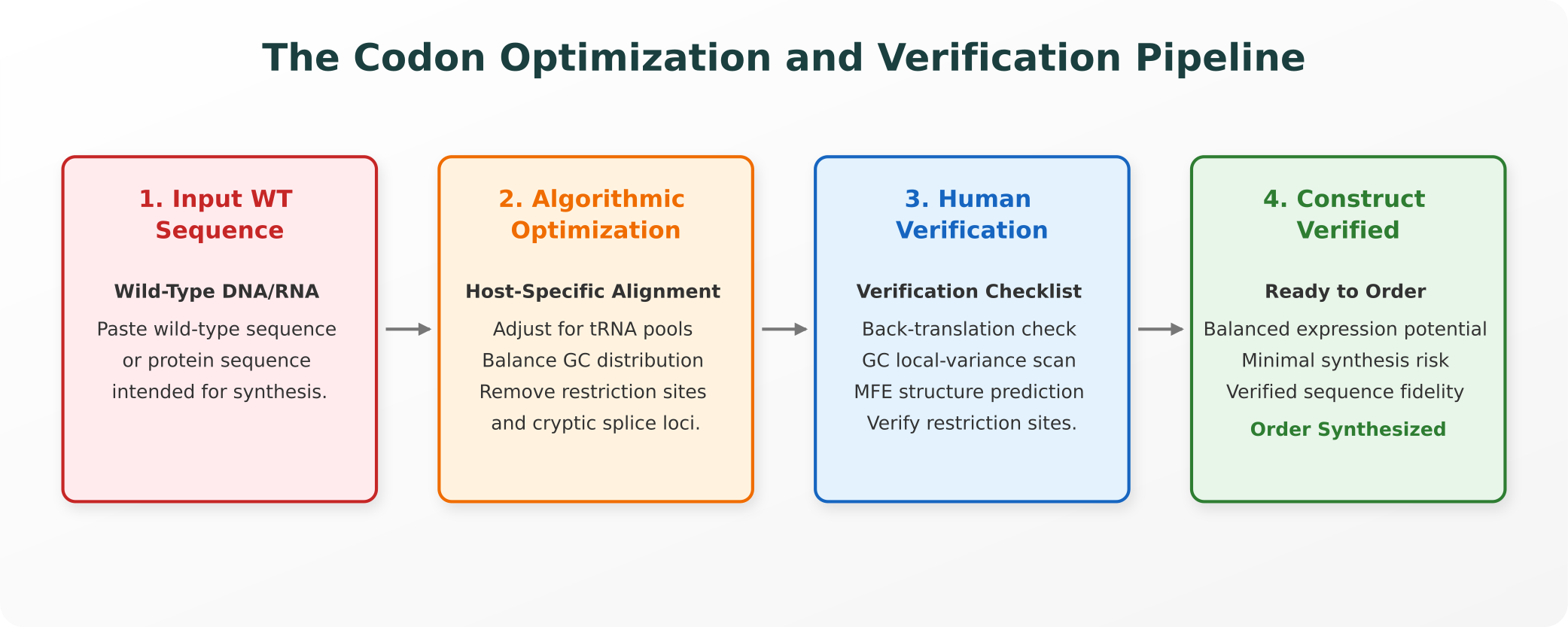
The Codon Optimization and Verification Pipeline
Before submitting a sequence for chemical synthesis, practitioners must guide their construct through a multi-stage alignment and verification check:
1. What Actually Changes Under the Hood?
Because the genetic code is redundant, multiple distinct codon triplets can translate into the identical amino acid. For example, six different codons encode for leucine. An optimization tool exploits this redundancy to alter several physical properties of your construct:
- Adjusting for Host tRNA Pools: Different species display distinct preferences for specific codons, a phenomenon known as codon usage bias. The tool calculates your sequence’s codon adaptation index (CAI) and systematically replaces rare codons with the “optimal” codons preferred by your expression host (such as Escherichia coli, yeast, or CHO cells). This aligns translation requirements with the host’s active tRNA pool, avoiding ribosomal stalling from depleted rare tRNAs.
- GC Content Alignment: The overall ratio of Guanine-Cytosine bases significantly impacts mRNA stability and transcription efficiency. Optimization algorithms align the GC percentage to match the host genome’s baseline (typically targeting a balanced 40% to 60% range).
- Removing Cryptic Sequences and Obstacles: The algorithm scans the sequence to eliminate unwanted motifs that can disrupt translation or transcription:
- Internal Restriction Sites: Removing sequences that match common restriction enzymes (like EcoRI or BamHI) to preserve downstream cloning options.
- Cryptic Splice Sites: Eliminating sequence patterns that eukaryotic splicing machinery might misinterpret, preventing unwanted truncation.
- Premature Poly-Adenylation Signals: Removing premature poly-A signals that can drive early termination in mammalian expression hosts.
2. What Optimization Does Not Guarantee
While online tools are marketed as reliable boosters of protein expression, the biological reality is far more complex. The literature indicates that codon optimization is not a guaranteed solution, and over-optimized sequences can underperform for several precise reasons:
- No Guaranteed Expression Increase: While optimizing codon adaptation index scores can relieve translation bottlenecks, it does not guarantee high expression yields. Published comparisons across diverse host systems indicate that the correlation between CAI and actual protein expression is highly variable, and some optimized genes show no improvement or even reduced yields compared to their wild-type counterparts.
- Altered mRNA Secondary Structures: Replacing codons changes the local nucleotide folding kinetics. If the optimization tool introduces highly stable hairpin loops near the ribosomal binding site, the resulting mRNA can fold into a tight conformation with high minimum free energy structure prediction values, blocking ribosomal entry and silencing expression.
- Stripping Out Critical Translational Pausing: Natural, wild-type sequences are not randomly organized. They frequently utilize rare codons at specific domain boundaries to induce deliberate, localized ribosomal pausing. This pausing is biologically critical: it grants the newly emerging peptide chain time to fold correctly before translation resumes. Replacing these rare codons with high-speed optimal codons can accelerate translation to the point where the emerging protein misfolds, aggregates, or becomes target-inactive.
3. Host-Specific Considerations
A sequence optimized for one organism will regularly fail or express poorly in another. The tRNA abundance profiles and GC preferences of E. coli are fundamentally different from those of mammalian cells.
For instance, E. coli highly prefers the GCG codon for alanine, whereas human cells prefer GCC. Applying a sequence optimized for bacterial synthesis directly in a mammalian assay can result in rapid ribosomal stalling, incomplete translation, and truncated protein products, highlighting why host-specific alignment is a non-negotiable requirement.
4. The Pre-Order Verification Checklist
Before committing institutional budgets and weeks of pipeline time to a synthesis order, researchers should perform several independent verification checks on their optimized sequence:
- The Back-Translation Check (Essential): Translate your optimized nucleotide sequence back into amino acids and run a pairwise alignment against your original wild-type protein sequence. Confirm that the sequence remains 100% identical. Even highly validated vendor algorithms occasionally introduce silent amino acid substitutions during restriction-site removal.
- Review Local GC Distribution: Do not rely on your construct’s average GC content score. Use sliding-window visualization tools to scan the local GC profile along the sequence. Look for localized GC-rich spikes (which can form highly stable, un-translatable hairpin structures) or AT-rich valleys (which can act as premature transcription termination signals), particularly near the 5’ end.
- Verify Restricting Sites: If you plan to insert your synthetic gene into a specific plasmid vector, manually scan the optimized sequence to confirm that none of your target restriction cloning sites have been introduced during the codon replacement process.
- Predict 5’ mRNA Folding Free Energy: Run a local minimum free energy structure prediction algorithm (such as ViennaRNA or mfold) specifically on the first 40 to 50 nucleotides of your mRNA sequence. Verify that the start codon region displays low structural stability (high, less-negative free energy values). Tight hairpin folds wrapping around the start codon or ribosomal binding site are the single most common cause of complete expression failure in synthetic constructs.
References and Authoritative Sources
For molecular biologists seeking to review the primary literature and biochemical datasets governing codon usage and synthetic gene design, the following publications serve as authoritative references:
- Codon Bias and Translation Kinetics: Plotkin, J. B., & Kudla, G. (2011). “Synonymous but not equivalent: the codes that shape gene expression and evolution.” Nature Reviews Genetics, 12(1), 32-42. doi:10.1038/nrg2899
- The Impact of mRNA Secondary Structure on Expression: Kudla, G. et al. (2009). “Coding-sequence determinants of gene expression in Escherichia coli.” Science, 324(5924), 255-258. doi:10.1126/science.1170160
- Translational Pausing and Protein Folding: Widmann, M. et al. (2020). “Effects of codon usage on folding of recombinant proteins.” Protein Expression and Purification, 165, 105486. doi:10.1016/j.pep.2019.105486
- Kazusa Codon Usage Database: The primary reference resource for genomic codon frequency tables across diverse species. kazusa.or.jp/codon
Explore Purna's Molecular Intelligence Platform
AI-powered workspace for biology teams to accelerate drug discovery from target identification to lead optimization.
Try Purna AI →
