
What "Validated Antibody" Actually Means on a Datasheet
For many wet-lab researchers, commercial antibodies are the workhorses of cellular and molecular characterization. To locate a target protein, map tissue localization, or perform a pulldown assay, scientists rely on commercial datasheets. Because these datasheets prominently display a green checkmark or the word "validated," practitioners frequently purchase and use these reagents without verifying their physical properties or validation parameters.
This trust is a significant, recurring source of reproducibility failures. In biological research, the word "validated" on an antibody datasheet is not a universal certificate of quality. It is a description of specific tests performed by the vendor under highly optimized, defined laboratory conditions. These conditions may have nothing to do with your target cell line, tissue structure, fixation protocol, or cellular abundance levels. Treating "validated" as a blanket guarantee frequently leads to non-specific binding, false-positive signals, and failed experiments that waste months of R&D resources.
This article serves as a definitive, technically precise reference guide to validated antibody datasheet records. We define what vendor validation actually covers, analyze the biophysical variables that cause these reagents to fail in new experimental contexts, and outline an independent verification workflow researchers should run before trusting an antibody for a new application.
What Vendor "Validation" Actually Covers
To evaluate the utility of an antibody datasheet, researchers must understand that vendor testing is strictly application-specific and highly optimized:
1. The Limits of Application-Specific Validation
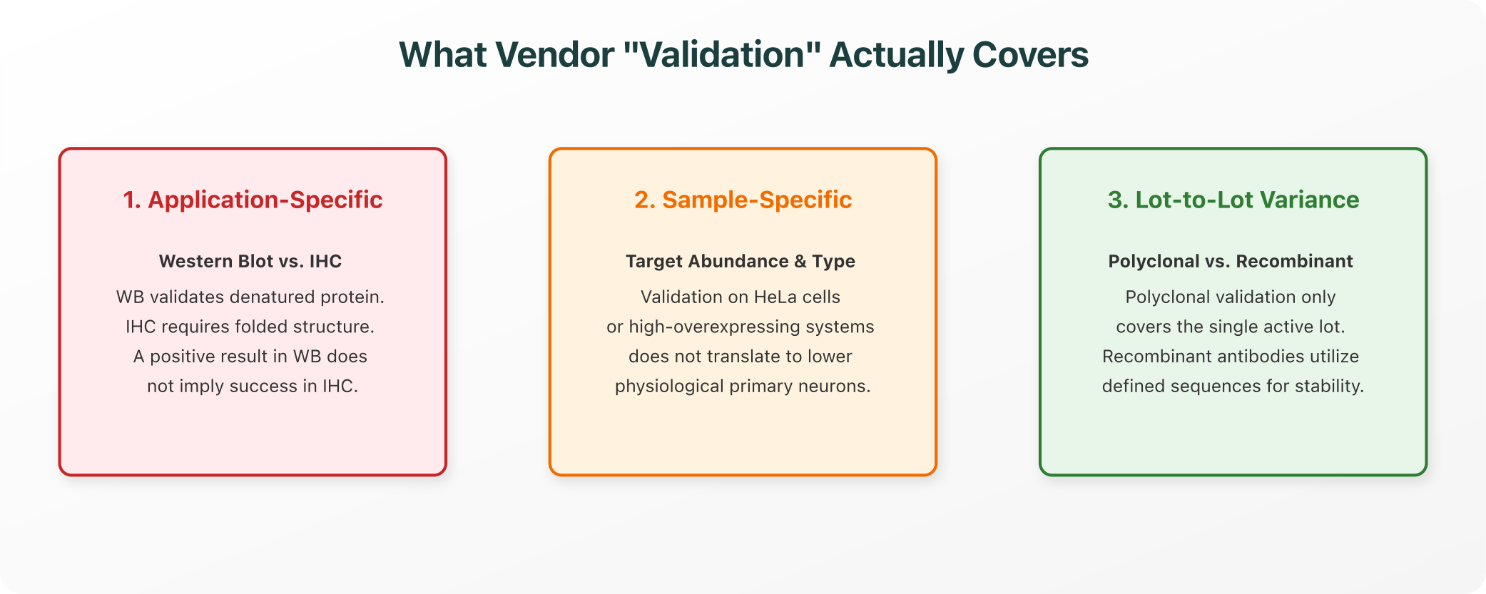
An antibody is never validated in the abstract: it is validated for a specific, defined application under specific conditions.
- Western Blot Validation (WB): A positive result in western blot validation means the antibody successfully bound its target protein after the sample was lysed, denatured, and reduced with SDS-PAGE. The target epitope is presented as a linear, unfolded peptide chain.
- Immunohistochemistry Validation (IHC): Conversely, IHC requires the antibody to bind its target in intact, folded 3D conformations within preserved tissue structures.
- The Disconnect: An antibody that successfully binds a denatured linear peptide in a Western Blot can completely fail to bind the native, folded protein in IHC because the target epitope is buried inside the 3D core. Conversely, an antibody validated for IHC can fail in WB because the denaturing process destroys the conformational epitope it recognizes.
- Sample-Specific Limitations: If an antibody is validated on HeLa cells (which highly overexpress the target), this does not mean it is validated for primary neurons, where the target is expressed at lower physiological levels. High expression can mask low-affinity, non-specific binding that becomes dominant when analyzing low-abundance primary samples.
2. Biophysical Variables That Drive Failure
Three main physical and chemical variables cause an antibody to fail when transitioned from the vendor's datasheet conditions to a new experimental pipeline:
- Species and Isoform Coverage: Many commercial antibodies are validated on human recombinant proteins or cell lines but fail on mouse or rat orthologs. Even minor evolutionary variations in the amino acid sequence can disrupt the specific epitope epitope binding pocket, neutralizing binding affinity.
- Fixation and Antigen Retrieval Sensitivity: Paraformaldehyde (PFA) or formalin fixation crosslinks proteins, structurally masking target epitopes. An antibody validated on native or frozen tissue often fails on fixed sections unless a specific antigen retrieval IHC protocol (such as heating the slide in an acidic buffer) is used to physically break the crosslinks and expose the hidden epitope.
- Sample-Specific Expression Levels: A positive result on a high-expressing cell line can fail on a sample where the target is expressed at lower physiological levels. High expression can mask low-affinity, non-specific binding that becomes dominant when analyzing low-abundance primary samples.
3. The Recombinant Antibody Distinction
To address systemic reproducibility issues, the international scientific community has driven a transition toward recombinant antibodies:
- Traditional Polyclonals: Polyclonal antibodies are harvested as raw sera from immunized animals. Because they contain a heterogeneous mixture of many different antibody clones targeting different epitopes, their specificity and binding affinity shift with every animal bleed, meaning validation on one lot tells you nothing about the specificity of the next lot.
- Traditional Hybridoma Monoclonals: While monoclonal, hybridoma cell lines can experience genetic drift, chromosomal loss, or contamination over multi-year storage cycles, resulting in lot-to-lot consistency issues.
- Recombinant Antibodies: Recombinant antibodies are produced by cloning the sequenced variable region genes (heavy and light chains) into defined expression vectors and expressing them in stable cell lines.
- The Impact on Reproducibility: Because the antibody sequence is genetically defined and registered, recombinant production completely eliminates lot-to-lot variance. In accordance with the IWGAV guidelines (International Working Group for Antibody Validation), recombinant antibodies offer the highest standard of reproducibility, ensuring that validation results remain stable across different production runs.
4. Pre-Experimental Verification Workflow
To prevent antibody-related failures, researchers should run a systematic, independent verification check before trusting any commercial datasheet:
- Review the Validation Parameters: Check if the vendor's validated applications (e.g., WB only) match your target application (e.g., IHC). If not, treat the antibody as unvalidated for your pipeline.
- Verify Species Homology (BLAST check): If you are working in a model organism (such as mouse) but the antibody was validated in human, locate the immunogen epitope peptide sequence on the datasheet. Run a pairwise BLAST alignment of the epitope against your target organism's ortholog. If the sequence conservation is below 90% or contains mutations in critical residues, the antibody is highly likely to fail or show reduced affinity.
- Establish Independent Controls: Never trust an experimental slide without proper positive and negative controls in your specific tissue/cell context:
- Positive Control: A cell line or tissue known to highly express the target.
- Negative Control: A knockout cell line (using CRISPR/siRNA), a tissue known to lack expression, or an isotype control antibody run on the identical sample.
- Consult Third-Party Databases: Cross-reference the antibody catalog number against independent curation databases (such as the Antibody Registry or CiteAb) to find peer-reviewed publications that used the exact same antibody in your target application and organism, verifying its actual field performance.
Closing: The Researcher’s Responsibility
For computational and molecular biology teams, the core take-away is clear:
- "Validated" is a historical description of what a vendor tested under highly optimized conditions.
- "Fidelity" is a claim about how an antibody performs in your specific experimental pipeline.
Treating the first as proof of the second is a fundamental error. Real recombinant antibody reproducibility requires researchers to take active responsibility for validation in their specific experimental context: verifying homology, checking epitope accessibility, and running proper positive and negative controls before trusting their results.
References and Authoritative Specifications
For researchers seeking to review the biochemical principles and validation protocols discussed, the following publications serve as authoritative references:
- IWGAV Guidelines for Antibody Validation: Landmark recommendations on validating antibodies across different applications. Uhlen, M. et al. (2016). "A proposal for validation of antibodies." Nature Methods, 13(10), 823-827. doi:10.1038/nmeth.3995
- Antibody Reproducibility Crisis: Baker, M. (2015). "Reproducibility crisis: Blame it on the antibodies." Nature, 521(7552), 274-276. doi:10.1038/521274a
- The Impact of Fixation on Epitope Accessibility: Bordeaux, J. et al. (2010). "Antibody validation." Biotechniques, 48(3), 197-209. doi:10.2144/000113382
- The Antibody Registry Database: Curation platform providing persistent, unique identifiers for commercial antibodies. antibodyregistry.org
Explore Purna's Molecular Intelligence Platform
AI-powered workspace for biology teams to accelerate drug discovery from target identification to lead optimization.
Try Purna AI →
