
What an Orthogonal Assay Actually Confirms, and Why One Assay Is Never Enough

A major, recurring source of attrition in early-stage drug discovery and target validation is the loose definition of assay replication. In many screening campaigns, the term “orthogonal” is used loosely to describe running an experiment again in duplicate, or using a different enzyme kit from a different manufacturer.
This conceptual shortcut is exceptionally dangerous. True orthogonality is not simply a matter of technical replication: it requires that the second assay evaluate the same underlying biological or physical phenomenon through a fundamentally distinct biochemical or biophysical mechanism. If your primary and secondary assays share the same detection modality, their potential sources of systematic error are identical, meaning you are merely repeating the identical method-specific artifacts.
This article serves as a definitive, technically precise reference guide to orthogonal assay workflows. We define what makes an assay genuinely orthogonal, analyze what a positive result across independent platforms does and does not prove, and explore standard examples of orthogonal confirmation in modern pharmacology.
The Orthogonal Assay Framework
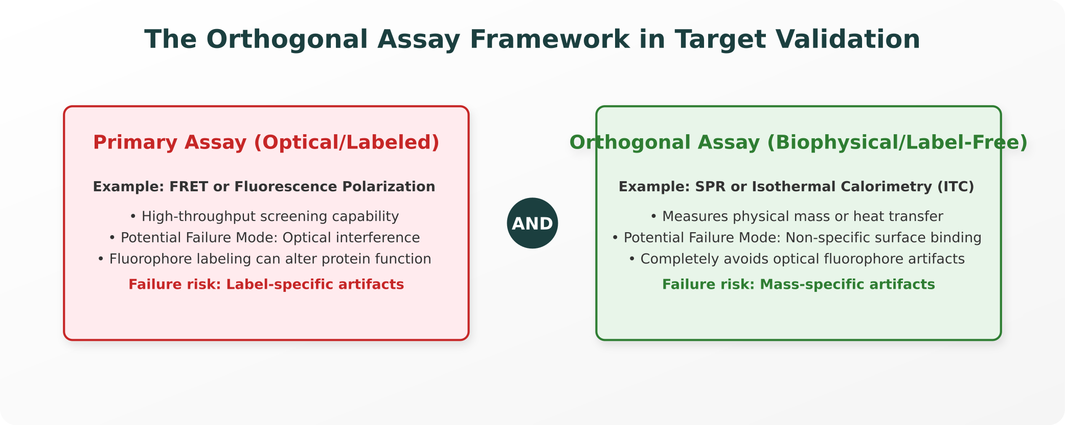
A rigorous validation campaign pairs complementary, independent detection modalities to ensure that candidate hits are not artifacts of a single method’s physical mechanism:
1. Defining Genuinely Orthogonal Assays
An assay is genuinely orthogonal to another when their potential sources of systematic error, physical limitations, and chemical artifacts are completely independent.
To understand this, consider the comparison of FRET versus SPR (Förster Resonance Energy Transfer versus Surface Plasmon Resonance) for confirming a protein-protein interaction:
- The Labeled Primary (FRET): FRET is an optical, fluorescence-based assay. It requires attaching fluorescent tags (donor and acceptor fluorophores) to your target proteins. A positive signal is generated when the proteins bind, bringing the fluorophores into close proximity.
- The Failure Mode: A fluorescent chemical compound from your screening library might autofluoresce or quench the signal, generating a false-positive reading. Additionally, the physical bulk of the fluorophore tag itself can structurally alter the protein, artificially forcing an interaction.
- The Label-Free Secondary (SPR): SPR is a biophysical, label-free assay. It measures the change in refractive index (mass) on a sensor surface when a ligand binds to an immobilized target.
- The Failure Mode: SPR does not use fluorescence, meaning fluorescent compounds cannot interfere with its readout. Its primary failure mode is non-specific binding of hydrophobic compounds to the sensor chip surface.
Because the physical artifacts that can corrupt a FRET reading (optical quenching) are completely unrelated to what can corrupt an SPR reading (non-specific mass accumulation), a candidate compound that scores positive in both assays is highly unlikely to be a method-specific artifact. This independent validation is what defines true orthogonality.
2. What Does Two-Assay Confirmation Actually Prove?
While scoring positive across two orthogonal assays is a critical milestone, researchers must remain epistemically cautious about what this result actually establishes:
- What It Establishes: It substantially reduces the mathematical probability that your hit is an artifact of your primary detection mechanism.
- What It Does Not Establish: It does not prove biological or therapeutic truth in a living organism. A compound can bind its target tightly in both a biochemical assay and a biophysical assay, yet fail to show any therapeutic efficacy in cellular or animal models due to poor membrane permeability or rapid degradation.
- The Shared Systematic Error (PAINS): Orthogonal biochemical assays cannot rule out compounds that act as Pan-Assay Interference Compounds (PAINS compounds). These molecules do not interact through specific, rational binding pockets. Instead, they form chemical aggregates, release reactive oxygen species, or physically chelate assay metals, generating false-positive signals across both optical and biophysical readouts. Identifying PAINS requires separate chemical validation assays, such as detergent-based counter-screens.
3. Common Cases of False Orthogonality
Many workflows that researchers assume are orthogonal are merely technical replicates with identical blind spots:
- Different Kits, Same Modality: Testing a binding hit using a second enzyme-linked immunosorbent assay (ELISA) kit from a different commercial vendor is not orthogonal. While it controls for vendor-specific batch defects, both assays still rely on antibody-antigen binding kinetics and enzymatic colorimetric readouts, leaving them equally vulnerable to non-specific antibody binding.
- Same Reporter, Different Cell Lines: Expressing the identical green fluorescent protein (GFP) reporter system in two different cell lines controls for cell-line specific genomic backgrounds, but it is not orthogonal. If your screening compound autofluoresces at the GFP emission wavelength, it will generate identical, false-positive readouts in both cell lines.
4. Concrete Examples in Target Validation
Modern, rigorous target discovery pipelines incorporate orthogonal steps as a standard requirement:
Target Engagement (Biochemical vs. Cellular)
Confirming that a small molecule physically engages its target inside a complex intracellular environment is a classic orthogonal challenge:
- The Primary: A biochemical binding assay (such as a thermal shift assay or fluorescence polarization) measures specific, direct binding between the purified protein and the ligand in a clean, cell-free buffer.
- The Orthogonal (CETSA): A CETSA target engagement (Cellular Thermal Shift Assay) evaluates this interaction directly inside living cells. CETSA exploits the physical principle that ligand-bound proteins are more thermally stable than unbound proteins. By heating intact cells to varying temperatures, lysing them, and quantifying the remaining soluble protein, researchers can verify target engagement in the presence of competing intracellular metabolites, confirming the drug actually crosses the cellular membrane to reach its target.
Genomic Perturbation (Independent Guides)
In functional genomics, confirming that knocking down a gene actually drives your target phenotype represents a different kind of orthogonality (independent perturbation, rather than independent detection):
- The Challenge: An siRNA or sgRNA sequence can induce an off-target knockdown of an unrelated gene, driving a false-positive phenotypic change.
- The Orthogonal Solution: To confirm the phenotype is real, researchers must replicate the effect using a second, completely non-overlapping sgRNA or siRNA sequence designed against a different region of the target gene. Because the two guides share zero sequence homology, they cannot share the identical off-target alignment profile. If both distinct guides produce the identical phenotype, the likelihood of an off-target artifact is effectively ruled out.
Closing: Independence of Failure Modes
To design rigorous, high-confidence validation campaigns, research teams must change the question they ask before placing synthesis or animal-model orders:
Do not simply ask: “Did we get a second positive data point?”
Instead, ask: “What biophysical or chemical failure would have to occur for both of these assays to give me the identical wrong answer?”
True orthogonality is defined by the absolute independence of failure modes. By pairing optical and biophysical, biochemical and cellular, or sequence-independent perturbation methods, computational and wet-lab teams can systematically expose method-specific artifacts, ensuring their R&D resources are directed only toward candidates of absolute biological validity.
References and Authoritative Specifications
For researchers seeking to review the biochemical principles and validation protocols discussed, the following publications serve as authoritative references:
- Assay Guidance Manual for Drug Discovery: Detailed guidelines on assay design, validation, and managing optical/chemical interference. ncbi.nlm.nih.gov/books/NBK53196
- PAINS Compounds and Interference: Baell, J. B., & Holloway, G. A. (2010). “New substructure filters for removal of pan assay interference compounds (PAINS) from screening libraries and for their exclusion in bioassays.” Journal of Medicinal Chemistry, 53(7), 2719-2740. doi:10.1021/jm901137a
- The CETSA Method: Martinez Molina, D. et al. (2013). “Monitoring drug-target engagement in cells and tissues using the cellular thermal shift assay.” Science, 341(6141), 84-87. doi:10.1126/science.1233606
- Biophysical Methods for Target Engagement: Holdgate, G. A. et al. (2019). “Biophysical methods in drug discovery.” Methods in Molecular Biology, 1888, 1-24. doi:10.1007/978-1-4939-8891-4_1
Explore Purna's Molecular Intelligence Platform
AI-powered workspace for biology teams to accelerate drug discovery from target identification to lead optimization.
Try Purna AI →