
What Is a Protein Embedding?
The scale and complexity of biological sequences have turned modern drug discovery into a high-dimensional data challenge. While traditional alignment-based methods have long served as the backbone of comparative genomics, they struggle to capture the complex, non-linear relationships that govern protein folding, structural dynamics, and biochemical function.
To bridge this gap, structural biologists and machine learning engineers are increasingly adopting protein embeddings as a unified numerical representation. Generated by a trained protein language model, these mathematical vectors translate the raw text of amino acid sequences into dense, continuous numerical spaces that capture the underlying physics and evolutionary history of proteins.
But what actually is a protein embedding, and how is it generated? This article defines the core mathematical concepts, explains the self-supervised training mechanisms behind state-of-the-art models like ESM-2, examines their unique Multiple Sequence Alignment (MSA) free advantage, and maps their citable downstream applications in protein embeddings drug discovery workflows.
From Sequence to High-Dimensional Space
The transformation of a biological sequence into a structured, high-dimensional vector space is a multi-step computational process:
1. Defining the Core Concept Precisely
At its most fundamental level, a protein embedding is a fixed-length, high-dimensional numerical vector representation of a protein sequence generated by a trained deep learning model.
- High-Dimensional Mapping: Instead of representing a protein as a string of categorical letters (such as "M-K-V"), the model projects each residue (and ultimately the entire sequence) into a dense vector space (typically consisting of hundreds or thousands of dimensions).
- Topological Conservation: The core mathematical property of a well-trained embedding space is that proteins with similar structural architectures or biological functions will land close to one another in that vector space (low Euclidean or cosine distance), even when they share no alignable sequence identity.
- Semantic Capture: This mapping is possible because the model's high-dimensional parameters capture the complex, non-linear rules of amino acid biochemistry (such as hydrophobicity, electrostatic charges, and steric constraints) directly from evolutionary data.
2. The Training Mechanism: ESM-2 Explained
To understand how these vectors are built, we can examine the evolutionary training pipeline of ESM-2 (Evolutionary Scale Modeling), developed by Meta AI:
Masked Language Modeling (MLM)
Like BERT in natural language processing, a protein language model is trained using a self-supervised approach known as Masked Language Modeling (MLM).
- Self-Supervision: The training process requires no manual labels or experimental structure data. Instead, the model is fed large-scale sequence databases derived from UniRef (such as UniRef50 and UniRef90), which span tens to hundreds of millions of diverse protein sequences.
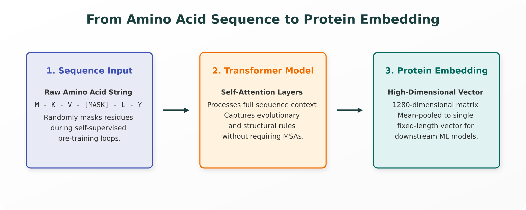
- The Training Loop: During each training epoch, a portion of the amino acids in each sequence are masked, following the masked language modeling convention popularized by BERT in natural language processing. The model must analyze the surrounding, unmasked sequence context and predict the exact identity of the hidden amino acid. By iteratively minimizing prediction error over millions of sequences, the model naturally learns the structural and evolutionary "grammar" of proteins.
The Transformer Architecture
ESM-2 utilizes a transformer-based, BERT-style encoder architecture:
- Self-Attention: Rather than processing sequences sequentially (like traditional recurrent neural networks), the transformer uses self-attention mechanisms to evaluate relationships between all amino acid pairs in a sequence simultaneously, regardless of their distance along the primary chain. This allows the model to capture long-range spatial contacts that are critical for folding.
- Scale and Parameterization: The ESM-2 family spans a wide range of model sizes, from 8 million to 15 billion parameters. In active research, the 650-million-parameter version is widely used. This model processes sequences across 33 self-attention layers, outputting a 1280-dimensional vector for each individual residue.
- Pooling: To generate a single representation for an entire protein, the per-residue vectors are typically "mean-pooled" (averaged across the sequence length) into a single, fixed-length 1280-dimensional vector.
3. The MSA-Free Advantage
Historically, predicting protein structure, function, or mutational tolerance required building a Multiple Sequence Alignment (MSA). By searching databases (such as UniProt) to find evolutionary relatives (homologs), researchers identify co-varying residues that physically suggest spatial contact.
While highly effective, the MSA approach has several structural limitations:
- High Computational Cost: Searching massive databases to align thousands of homologous sequences for a single query protein is computationally slow and resource-intensive.
- The Orphan Bottleneck: If a target protein is novel, highly engineered, or belongs to an understudied organism (such as viral or eukaryotic proteins with few known evolutionary relatives), the MSA will be shallow or noisy, rendering downstream structure and function predictions highly unreliable.
Protein language model embeddings completely bypass this bottleneck. Because the transformer has already encoded the global rules of protein biochemistry within its parameters during pre-training, it can generate high-fidelity structural and evolutionary representations directly from a single sequence, with no MSA search required. This provides a massive speed and accuracy advantage when working with poorly characterized orphan proteins.
4. Downstream Applications in Drug Discovery
Rather than remaining theoretical abstractions, these high-dimensional vectors serve as the primary feature input for several active, published research applications:
- Protein-Ligand Binding Affinity: Machine learning models use protein embeddings combined with small-molecule embeddings to predict binding affinity ($K_d$, $K_i$) and screen massive chemical libraries for novel inhibitors.
- Protein-Protein Interaction (PPI) Prediction: By calculating distance vectors between distinct protein embeddings, models can predict whether two proteins physically interact in a cellular pathway, mapping complex interactomes.
- Drug-Target Interaction (DTI) Mapping: Neural networks leverage embeddings to screen approved compound libraries against novel targets, prioritizing candidates for active computational drug repurposing.
- Residue-Residue Contact and Structural Prediction: As demonstrated by models like ESMFold, the internal attention patterns of mid-to-late transformer layers can be directly decoded to predict 3D coordinate structures with experimental-grade accuracy, entirely bypassing the need for MSA pipelines.
5. Ongoing Research Nuance: Disentangling Embeddings
A key area of ongoing, sophisticated research in structural bioinformatics is interpretability. For years, embeddings were treated as "black boxes" (high-dimensional vectors that worked well for machine learning but remained uninterpretable to human scientists).
Recent research has uncovered two critical nuances:
- Layer-Specific Specialization: Rather than presenting a clean, settled division of labor across the network, interpretability is an active and developing field of study. Probing studies have found that mid-to-late transformer layers (in one study, specifically layers 20 through 33 of a 33-layer model) capture richer function-level and motif-level information than the final layer alone, showing that the representation of biological concepts within the network is highly distributed and nuanced.
- Sparse Autoencoders (SAEs): Researchers are increasingly using sparse autoencoders to "disentangle" these dense embeddings. By projecting the 1280-dimensional vector into a sparser, higher-dimensional space, they can identify individual activation features that correspond to real biological concepts, such as kinase active-sites, zinc fingers, or transmembrane regions. This active area of research promises to make AI-driven biological design fully transparent and interpretable.
6. Honest Limitations
To maintain scientific integrity, researchers must recognize the boundaries of language-model-based representations:
- Statistical Patterns, Not Physical Laws: Protein embeddings represent learned statistical probabilities from UniRef databases, not direct physical or thermodynamic measurements. They do not simulate active molecular dynamics or capture real-time conformational changes.
- The Distribution Barrier: If a protein sequence is highly engineered, synthetic, or lies completely outside the evolutionary distribution of the UniRef training data, the generated embedding may be inaccurate or misleading.
- Mandatory Experimental Validation: Any downstream target or binding affinity predicted via protein embeddings must be treated as a computational hypothesis. Strict wet-lab validation (such as in vitro binding assays and crystallographic validation) remains a non-negotiable requirement for clinical translation.
Purna AI: Grounding Embeddings in Hard Biochemistry
Generating and processing protein embeddings typically requires extensive computational infrastructure, command-line coding, and manual tool-switching between Python notebooks and structural viewers.
Purna AI's Molecular Intelligence Platform (MIP) integrates these advanced machine learning models natively into a single, cohesive environment:
- Direct MSA-Free Structural Prediction: Purna integrates ESMFold directly into your workspace. You can generate accurate 3D structures from single amino acid sequences in seconds with zero configuration or MSA pipeline building.
- Interactive 3D Visualization: Purna renders predicted structures natively using Molstar. You can instantly overlay embeddings-derived contact maps, color residues by model confidence, and inspect active pockets.
- Evidence-Backed Annotation: Purna does not leave your computational models in isolation. Every target, domain, and variant identified via embeddings is automatically cross-referenced in real-time with 30+ live biological databases (ClinVar, UniProt, OMIM), delivering cited, factual summaries with clickable provenance.
By combining advanced protein language modeling with rigid physical and clinical grounding, Purna AI transforms high-dimensional machine learning into precise, evidence-backed molecular intelligence, allowing your research teams to design therapies with absolute scientific integrity.
Explore Purna's Molecular Intelligence Platform
AI-powered workspace for biology teams to accelerate drug discovery from target identification to lead optimization.
Try Purna AI →