
How Scientists Actually Diagnose a Disease at the Molecular Level

For generations, the process of diagnosing a disease was primarily observational. Doctors relied on visible symptoms, patient-reported histories, physical imaging, or basic chemical measurements (such as blood glucose or cholesterol levels) to infer what was happening inside the body. While highly effective for many common conditions, this top-down approach has limitations when dealing with complex, genetically-driven, or infectious diseases.
Today, medicine is undergoing a profound transition from observational to molecular. Known as molecular diagnosis, this approach allows scientists to look directly at the biological code of life: DNA, RNA, and specific biomarker molecules extracted from a patient sample. By analyzing these microscopic signatures, clinicians can identify the precise root cause of a disease, often long before visible symptoms ever appear.
This article provides an accessible, evidence-grounded explainer on how diseases are diagnosed at the molecular level, demystifying the core laboratory techniques used, illustrating their utility with real-world clinical examples, and outlining the critical role of data interpretation in diagnostic accuracy.
The Journey From Sample to Diagnosis
Translating a raw patient sample into a precise molecular signature is a highly structured process:
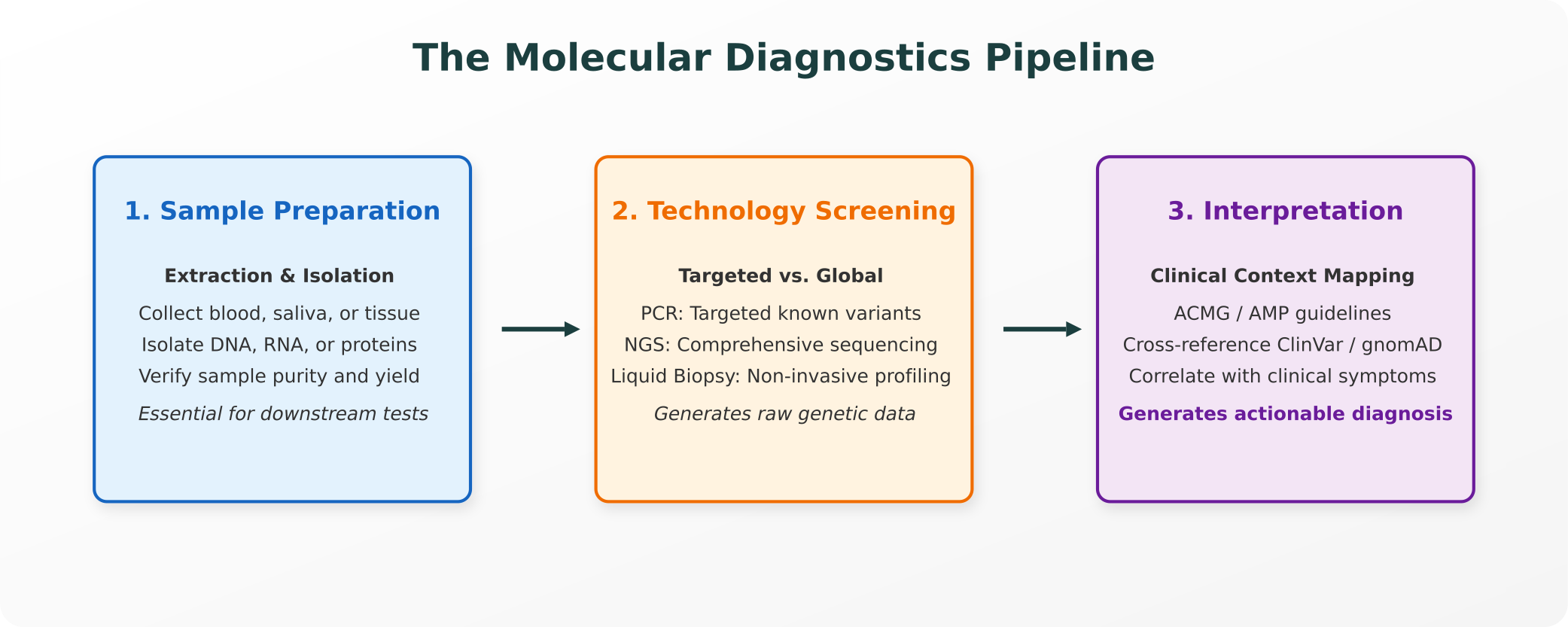
1. The Core Technique Toolkit
To construct a molecular profile, diagnostic laboratories utilize a specialized set of molecular diagnostics techniques, each optimized for distinct clinical scenarios:
PCR (Polymerase Chain Reaction)
PCR is the workhorse of molecular diagnostics. It is used when scientists need to detect the presence or absence of a specific, pre-determined genetic sequence with high speed and sensitivity.
- The Mechanism: PCR acts as a molecular photocopying machine, taking a tiny fragment of target DNA and amplifying it millions of times so it can be detected.
- Digital PCR (dPCR) and Droplet Digital PCR (ddPCR): These advanced techniques partition a single sample into thousands of individual, microscopic nanoliter droplets. Because each droplet undergoes an isolated reaction, scientists can achieve absolute, precise quantification of rare genetic mutations. This is incredibly useful for tracking a known genetic signature (such as cancer mutations in circulation) over time.
NGS (Next-Generation Sequencing)
While PCR checks for specific, known targets, NGS is used when researchers need a broader genomic view.
- The Mechanism: NGS sequences millions of DNA fragments in parallel. Instead of checking a single spot, it can profile hundreds of genes simultaneously or sequence a patient’s entire exome or genome, allowing clinicians to discover unknown variants or map complex genetic conditions.
Microarray Analysis
Microarrays are solid surfaces (such as glass slides) printed with thousands of microscopic spots, each containing a specific DNA probe.
- The Mechanism: When a patient’s sample is applied to the slide, complementary sequences bind (hybridize) to the probes, emitting a fluorescent signal. This allows for massive, multiplexed profiling of gene expression or variant detection in a single, high-throughput assay.
Liquid Biopsy
Traditionally, diagnosing deep-tissue diseases (like cancer) required an invasive surgical biopsy.
- The Mechanism: Liquid biopsy is a non-invasive alternative that extracts and analyzes circulating tumor DNA (ctDNA) or other disease-relevant molecular material directly from a simple blood draw. While not a complete replacement for traditional tissue biopsy, it serves as an invaluable complement, particularly for monitoring minimal residual disease (MRD) and tracking treatment response over time.
2. Real-World Clinical Applications
These diagnostic techniques are actively applied across multiple areas of medicine, directly guiding clinical decision-making:
- Infectious Disease: PCR is the gold standard for rapid pathogen detection, such as identifying the SARS-CoV-2 virus. Beyond simple detection, NGS is increasingly used to sequence bacterial pathogens (such as Mycobacterium tuberculosis), allowing clinicians to map specific antimicrobial resistance genes and select the most effective antibiotic cocktail.
- Oncology: Targeted cancer therapies are highly specific. For example, in non-small cell lung cancer, clinicians use NGS to check for specific mutations in the EGFR (epidermal growth factor receptor) gene. Identifying these molecular signatures allows oncologists to bypass standard chemotherapy in favor of targeted tyrosine kinase inhibitors, improving patient outcomes.
- Inherited/Genetic Diseases: Prospective parents utilize genetic testing for disease to undergo carrier screening for conditions like cystic fibrosis. This allows families to understand their risk profiles and make informed reproductive decisions based on precise molecular evidence.
- Pharmacogenomics: Our genetic makeup dictates how we process medications. Clinicians test for variants in the CYP2C19 gene (which encodes a key liver enzyme) to guide dosing decisions for clopidogrel, a blood thinner. Patients with loss-of-function variants convert the drug to its active form significantly less efficiently, which can affect both treatment efficacy and safety, making molecular profiling clinically important.
3. The Interpretation Boundary: Data is Not a Diagnosis
A common misconception is that generating a genetic sequence or molecular measurement is equivalent to a diagnosis. In reality, raw laboratory data is merely the starting point.
The Role of Clinical Interpretation
When NGS identifies a genomic variant, clinicians must determine what that variant actually means for the patient.
- ACMG/AMP Guidelines: Laboratories use strict, evidence-based frameworks established by the American College of Medical Genetics and Genomics (ACMG) and the Association for Molecular Pathology (AMP) to classify variants into five categories: pathogenic, likely pathogenic, variant of uncertain significance (VUS), likely benign, or benign.
- Evidence Cross-Referencing: Scientists must cross-reference detected variants against curated global databases (such as ClinVar and gnomAD) and peer-reviewed literature to see if the variant has been observed in other patient populations and what phenotypes are reported.
- The Diagnostic Equation: A molecular test is never interpreted in isolation. Diagnostic performance depends heavily on sample collection quality, laboratory workflow validation, and the patient’s active clinical presentation.
4. Honest Limitations of Molecular Testing
While incredibly powerful, molecular diagnostics are not infallible:
- Sensitivity and Specificity Limits: No molecular test is perfectly sensitive (capable of detecting 100% of true cases) or perfectly specific (free of all false positives). Contamination, low-yield extractions, or genetic mosaicism can result in false negatives.
- Complementary Nature: Liquid biopsy, despite its non-invasive appeal, remains a complement to tissue biopsy in most oncology contexts. If a liquid biopsy returns a negative result, a traditional tissue biopsy is still required to rule out the presence of a localized tumor.
- Contextual Correlation: An abnormal molecular marker does not automatically equal disease. For example, carrying a genetic risk factor does not guarantee that an individual will ever develop the associated condition (variable penetrance), meaning clinical correlation remains a fundamental requirement.
Purna AI: Enabling Evidence-Grounded Diagnostic Curation
Manually analyzing a molecular report requires researchers and clinicians to jump between different visualizers, genomic databases, and literature browsers.
Purna AI’s Molecular Intelligence Platform (MIP) is designed to eliminate this fragmented curation loop, uniting data and evidence in a single workspace:
- Variant Classification and Grounding: Purna automates the tedious steps of variant annotation. When a genomic variant is queried, Purna instantly cross-references ClinVar, gnomAD, and UniProt in real-time, delivering a structured summary of clinical significance, allele frequencies, and functional domains.
- 3D Structural Interpretation: Purna integrates molecular biology directly into the diagnostics workspace. Using Molstar, clinicians can visualize mutated proteins in 3D, inspect active-site structural changes, and run biophysical simulations to evaluate how a mutation impacts folding stability ($\Delta\Delta G$).
- Factual Provenance: Purna does not write ungrounded text. Every biological mechanism, clinical association, or literature summary generated by the platform is anchored directly to live reference databases with clickable, verified citations.
By integrating molecular datasets, structural visualization, and literature curation into a single biology IDE, Purna AI transforms raw genetic data into precise, evidence-backed molecular intelligence, empowering research and clinical teams to make decisions with absolute scientific integrity.
Explore Purna's Molecular Intelligence Platform
AI-powered workspace for biology teams to accelerate drug discovery from target identification to lead optimization.
Try Purna AI →